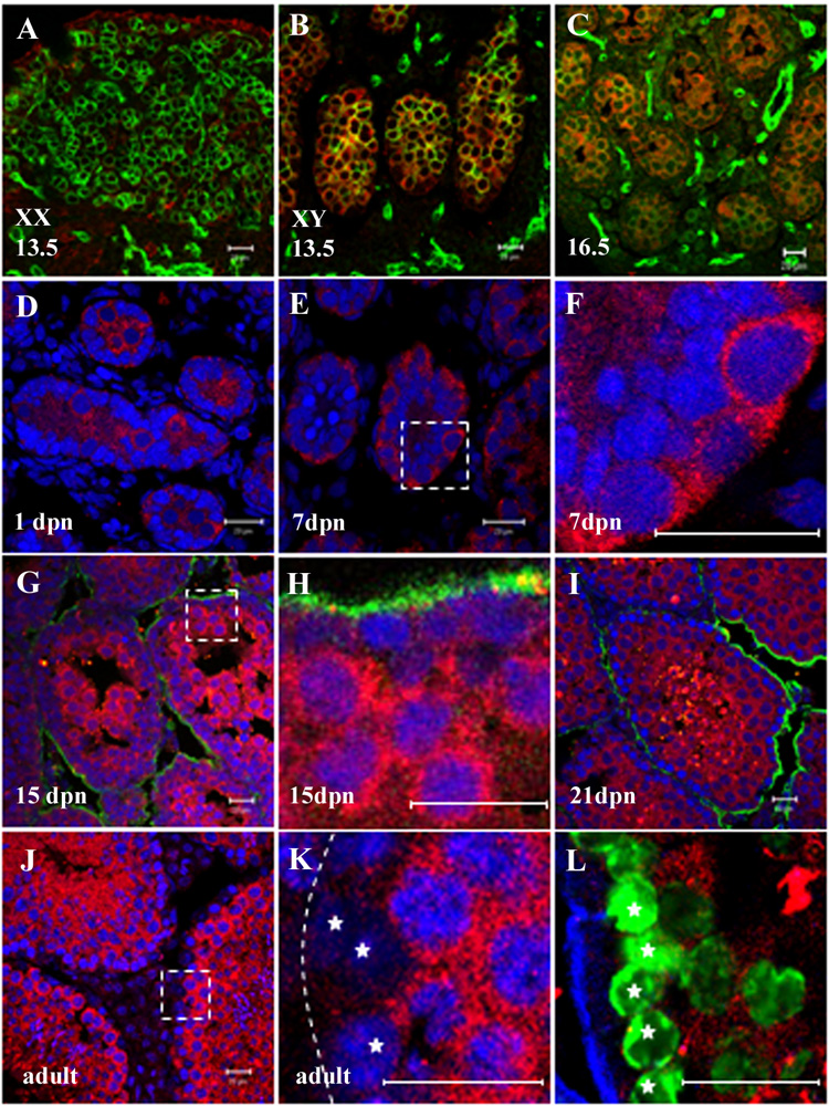
Figure 4. GM114 is a cytoplasmic protein expressed in male germ cells.

Fluorescent immunostaining was performed on gonads using a polyclonal antibody against GM114 (red) during fetal stages (A–C) and in postnatal and adult testes (D–L). (F), (H), and (K) are higher magnification of boxed regions in (E), (G) and (J). GM114 protein was detected in the cytoplasm of XY germ cells at 13.5 dpc (B) and16.5 dpc (C), but not in XX germ cells (A). A PECAM antibody labels germ cells and vascular endothelial cells (green in A–C). GM114 is maintained in all male germ cells from 1 to 7 dpn (D–F). At 15 dpn GM114 expression remains high in differentiated spermatocytes in the center of the seminiferous cords, but has declined in undifferentiated spermatogonia at the cord periphery (G, H). This pattern is seen at all later stages (J–L). Undifferentiated spermatogonia (stars in K and L, with low DNA density) have no or very low expression of GM114 compared to high expression in adjacent spermatocytes (high DNA density). The identity of undifferentiated spermatogoina at the periphery of the cord was confirmed by labeling with GCNA1 antibody (green in K) which marks spermatogonia and early spermatocytes (stars). DNA was stained with propidium iodide (blue in D–K). Basal lamina outlining testis cords is stained with an antibody against laminin (green in G–I; blue in L), or designated by a dashed line in K. All bars represent 20µm.
