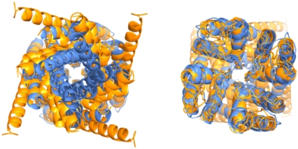
FIGURE 1.
Superposition of pore domains of potassium channels of known structure as seen from the intracellular (left) and extracellular (right) ends using a ribbon representation. Open (orange) forms include the MthK, KvAP, and Shaker structures, and closed (blue) forms include the KcsA, KirBac 1.1, and NaK structures. Orientation of each channel was accomplished by least-squares fitting each structure onto KcsA using the Cα atoms ranging from the pore helix to glycine hinge on the S6 inner helix.

