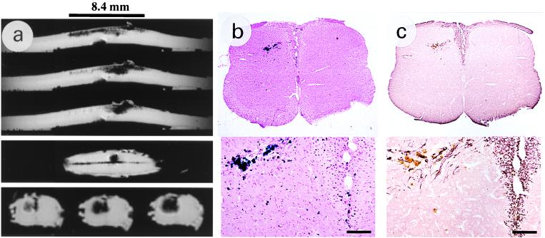
Figure 3.
Md rat 10 days after transplantation of magnetically labeled CG-4 cells. (a) Shown are the three MRI planes of view for TE = 6 msec at 78 μm isotropic resolution. In the sagittal plane (top images, consecutive slices), cellular migration can be appreciated over a distance of 8.4 mm. The contrast in the transverse images (enlarged in bottom row, shown is each third interleaved slice) corresponds to the area of Prussian blue staining (b Upper) and antiproteolipid protein immunolabeling (c Upper) and shows a blooming effect caused by an extended-range susceptibility effect of the magnetic particles. (b and c Lower) Cell migration from the injection site toward the dorsal column, where the majority of the newly formed myelin was found. Note the differential orientation of the individual myelin fibers, which is along the direction of CG-4 cell migration. (Bars represent 100 μm in b and c Lower.)

