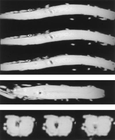
Figure 5.
Md rat 14 days after transplantation of magnetically labeled and paraformaldehyde-fixed (dead) CG-4 cells. Shown are the three MRI planes of view for TE = 6 msec, with consecutive slices for the sagittal plane (top images) and interleaved slices for the transverse plane (bottom row, enlarged). In this control experiment, contrast is visible at the injection site only; no cellular migration can be observed. Magnifications: ×4, Top and Middle.

