Figure 1.
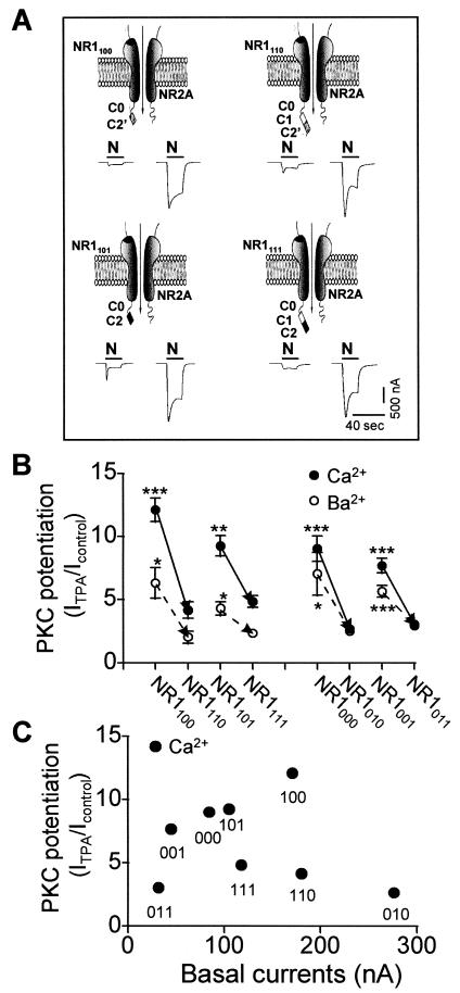
Splicing in of the C1 cassette reduces PKC potentiation. NMDA responses recorded from Xenopus oocytes expressing NR1/NR2A receptors before and after treatment with TPA at a holding potential of −60 mV. (A) Currents recorded in Ca2+ Ringer's solution before (Left) and after (Right) treatment with TPA (100 nM; 10 min) from oocytes expressing four receptors: NR1100/NR2A, NR1110/NR2A, NR1101/NR2A, and NR1111/NR2A. The rows show receptors that differ by presence or absence of the C1 cassette; columns show receptors that differ by presence of C2′ vs. C2. (B) Mean PKC potentiation values for eight receptors arranged in pairs; arrows show the effect of splicing in of C1. C1-containing receptors showed reduced PKC potentiation (filled circles, Ca2+ Ringer's solution; open circles, Ba2+ Ringer's solution). For NR1100/NR2A vs. NR1110/NR2A, potentiation was to 12.1 ± 1.0 (n = 11) vs. 4.2 ± 0.7 (n = 6) times the control response in Ca2+ (P < 0.001; Upper Right vs. Upper Left records in A). For NR1101/NR2A vs. NR1111/NR2A, potentiation was to 9.3 ± 0.9 (n = 8) vs. 4.8 ± 0.5 (n = 5) times the control response (P < 0.01; Lower Right vs. Lower Left records in A). *, P < 0.05; **, P < 0.01; ***, P < 0.001. (C) The degree of PKC potentiation is not inversely related to basal current.

