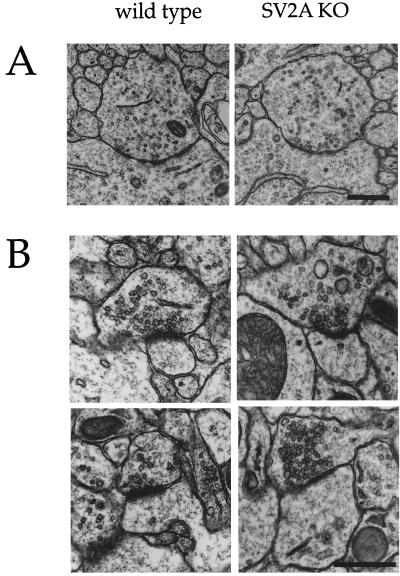
Figure 6.
Synapse morphology is normal in SV2A-knockout mice. (A) Shown are representative electron micrographs of symmetric (GABAergic) synapses in the cell-body region of CA3 hippocampal neurons. These synapses are presumed to be GABAergic based on their location, symmetry of active zones, and irregularly shaped vesicles. Qualitative inspection of these synapses suggested no difference in density or morphology between wild type and SV2A knockouts. (B) Shown are representative electron micrographs of asymmetric synapses onto proximal dendrites of CA3 hippocampal neurons. Synapses in this region are primarily from dentate granule cells and are largely asymmetric, presumed glutamatergic synapses. Similar morphology was observed in wild type and SV2A knockouts. Quantitative analyses of these synapses are presented in Table 1.

