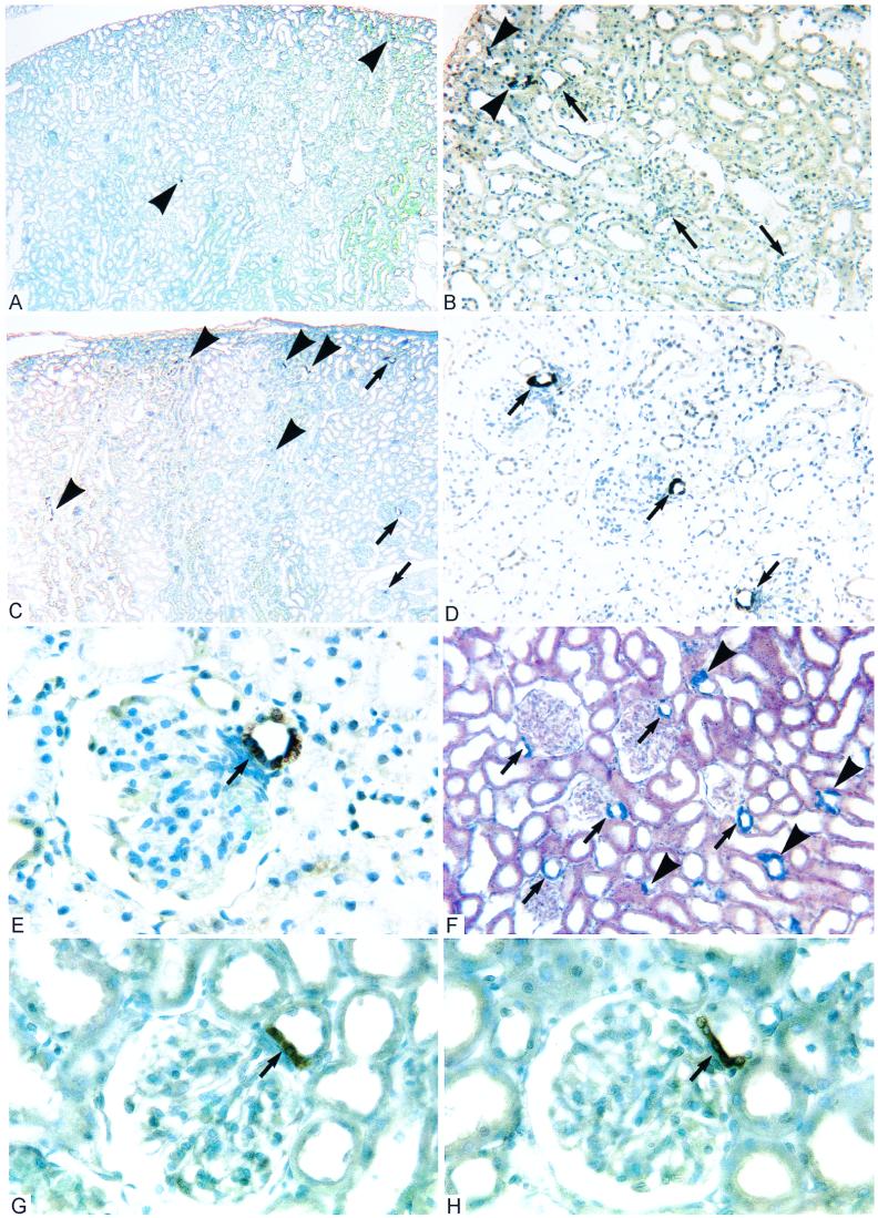
Figure 1.
Histological sections of kidney cortex from adult rats (>250 g). (A and B) After ADX combined with CS replacement for 2 weeks, intense COX-2-ir is observed in macrophages (▸) and a very few scattered cTAL cells, but generally is absent from the macula densa (➞). These images are indistinguishable from controls. (C and D) ADX littermate without CS replacement shows intense COX-2-ir in some cTAL (▸) and nearly all macula densa (➞). (E) At higher magnification, it is apparent that COX-2-ir fills the cytoplasm of macula densa cells (➞) as well as cTAL cells in the opposite wall. (F) In situ hybridization of ADX kidney demonstrates COX-2 mRNA at sites identical to COX-2-ir. (G and H) COX-2 expression is observed at the macula densa (➞) in control rats administered RU486 (G) or spironolactone (H) to block steroid receptors (GR and MR, respectively). (Figure widths: A and C, 2.4 mm; B, D, and F, 600 μm; E, G, and H = 180 μm.)

