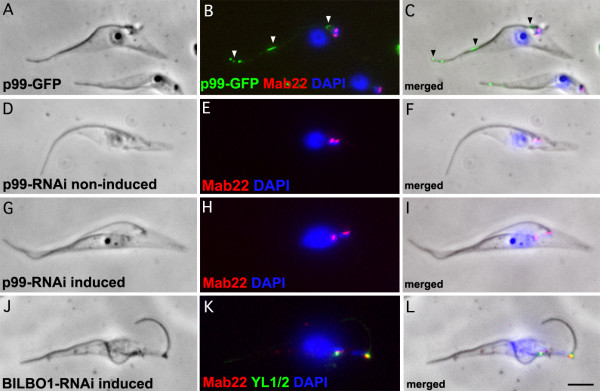
Figure 3.
GFP tagged and immunofluorescence on RNAi knockdown cells. A-C. 48 h induced p99-GFP expressing cytoskeletons (green) probed with Mab 22 (red) and DAPI (Blue). We made a GFP tagged protein because the rabbit antibody raised to the 99 kDa was positive by Western blots on E. coli expressed purified proteins and trypanosome cells but was not detectible by immunofluorescence. D-F. Non-induced p99 RNAi cytoskeletons probed with Mab 22 and DAPI (Blue). G-I. 48 h induced p99 RNAi cytoskeletons probed by Mab 22 and DAPI (Blue). K-L. BILBO1 24 h induced RNAi cytoskeletons probed with Mab 22 and YL1/2 and DAPI (Blue). Note that the Mab 22 antibody signal remains present after probing p99 and BILBO1 RNAi induced knockdown cytoskeletons indicating that the gene expressing the TAC protein is neither p99 nor BILBO1. In A-L scale bar is 5 μm.

