Figure 2.

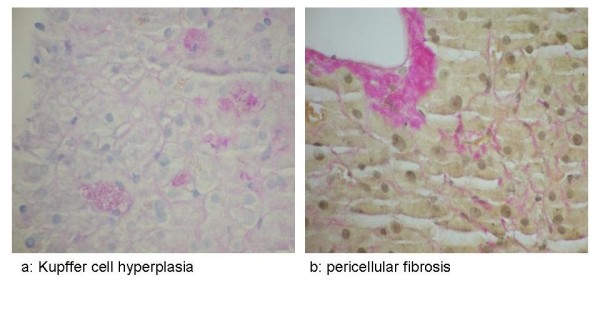
PAS diastase and Van Gieson stains. (a) PAS diastase stain showing Kupffer cell hyperplasia. (b) Van Gieson stain showing pericellular fibrosis adjacent to a terminal hepatic venule.

PAS diastase and Van Gieson stains. (a) PAS diastase stain showing Kupffer cell hyperplasia. (b) Van Gieson stain showing pericellular fibrosis adjacent to a terminal hepatic venule.