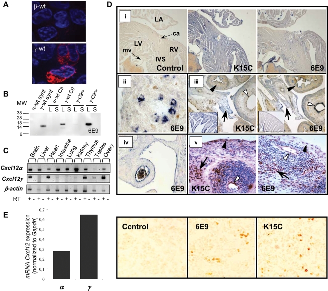
Figure 1. Tissue expression of γ-wt in human and mouse.
(A) Specific immunofluorescent detection of γ-wt. HEK-293T cells were transfected either with β-wt- (upper panel) or γ-wt- (lower panel) expressing pCDNA3.1 plasmids, treated with Brefeldin A, permeabilised with saponin and labelled with the 6E9 mAb and a Texas Red anti-mouse IgG. The nuclei of cells were counterstained with DAPI. Images are representative of six independent determinations. Original magnification ×63. (B) Mutagenesis of K78/K80 in γ-wt C9 (γ-C9up) prevents the specific recognition of the γ-wt chemokine by the 6E9 mAb. Western blot analysis of chemically synthesized α-wt and γ-wt (synt) and α-wt C9, γ-wt C9, γ-C9up and γ-C9dw C9-tagged chemokines expressed from SFV-vectors in BHK cells. Cell lysates (L) or culture supernatants (S) were separated by SDS-PAGE and probed with 6E9 mAb and a HRP-sheep anti-mouse Ig secondary antibody. MW, molecular weight in KDalton. Results are representative of two independent determinations. (C) Expression of Cxcl12α and Cxcl12γ mRNAs by RT-PCR in different adult mouse tissues. β-actin was used as loading control. RT +/− denotes presence or absence of RT enzyme. Data are representative of three independent determinations. (D) Detection of CXCL12 isoforms either with K15C mAb or anti-γ-wt 6E9 mAb in mouse and human tissues. (i) Mouse adult heart. LA, left auricle; LV, left ventricle; RV, right ventricle; IVS, interventricular septum; ca, carotid artery; mv, mitral valve. (ii) Detail of a lung bronchiol (mouse E16.5 embryo). (iii) Mouse E16.5 embryo intestin and bladder. White arrowheads, bladder epithelium; black arrowheads, large intestine; arrows, peritoneum. In inset, details of intestinal mucosa labeling. (iv) Large abdominal vessel (mouse E16.5 embryo). (v) Human inflammatory synovial tissue (rheumatoid arthritis). White arrowheads, blood vessel; black arrowheads, lining synoviocytes; arrows, fibroblasts. Control: secondary antibody. Original magnifications ×4 (i,iii inset), ×10 (iii), ×20 (iv), ×40 (ii) and ×400 (v). (E). CXCL12 expression in the mouse bone marrow. Left panel, expression of Cxcl12α (α) and Cxcl12γ (γ) mRNAs determined by quantitative real time-PCR and normalized to Gapdh expression. Results are representative from three independent determinations for each PCR reaction. Right panel, detection of CXCL12 isoforms by use of either K15C or anti-γ-wt 6E9 mAb. Control: secondary antibody. Original magnification (×40).

