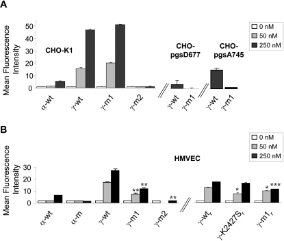
Figure 3. Cell surface GAG-binding activity of α-wt and γ-wt.
(A) Parental (K1) or GAG-mutant (pgsD677, pgsA745) CHO cells were incubated with the indicated concentration of wt (α-wt, γ-wt) or mutant (γ-m1, γ-m2) chemokines for 60 min at 4°C, and after extensive washing to remove free chemokine, were labelled with K15C mAb and a PE-goat anti-mouse Ig secondary antibody. Fixed cells were analyzed by flow cytometry. Values represent the mean fluorescence intensity±SD of three independent experiments performed in triplicate (B) Primary human-microvascular endothelial cells (HMVEC) were incubated with the indicated concentration of chemically synthesized (α-wt, α-m, γ-wt, γ-m1, γ-m2; left) or recombinant (γ-wtr, γ-K2427Sr, γ-m1r, right) chemokines and treated as in (A). Values represent the mean fluorescence intensity±SD of two independent experiments performed in triplicate. *p<0.05, **p<0.01, ***p<0.005 as compared to the binding obtained for the corresponding concentration of γ-wt (for γ-m1 and γ-m2) or γ-wtr (for γ-k2427Sr and γ-m1r) chemokines.

