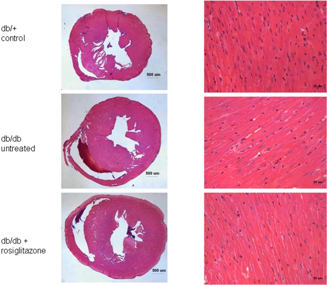
Figure 2. High-power light microscopy slides of myocardial tissue from rosiglitazone-treated and untreated db/db mice, and db/+ controls.
Individual cardiomyocytes from all three groups appear qualitatively similar based on morphology and size, though the untreated db/db cardiomyocytes exhibit very mild thickening. No increases in inflammatory cells, cell death, fibrosis, or other processes were observed in the two db/db groups compared to db/+ controls.

