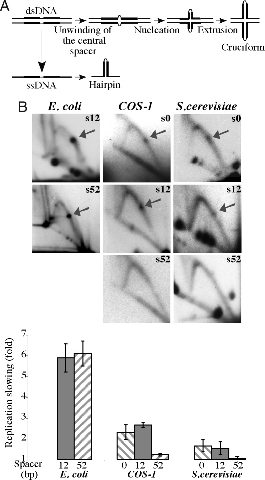
Fig. 4.
Replication fork stalling at inverted Alu repeats with varying lengths of the central spacer. (A) Schematic representation of hairpin and cruciform structure formation by an IR. (B) 2D gels and quantitative analysis of replication stalling at Alu IRs with 100% sequence homology and either 0 bp (s0), 12 bp (s12) or 52 bp (s52) spacers in E. coli, COS-1 cells, and S. cerevisiae

