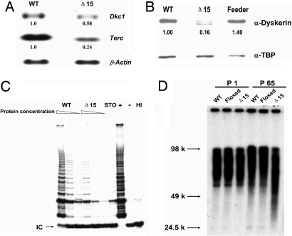
Fig. 1.
Decreased levels of Terc RNA and accelerated telomere shortening in Dkc1Δ15 ES cells. (A) Decreased levels of Terc RNA in Dkc1Δ15 ES cells as assessed by Northern blot analysis. Ten micrograms of total RNA from WT and Dkc1Δ15 (Δ15) ES cells were loaded in each lane and hybridized sequentially with probes for Terc, Dkc1, and β-actin. (B) Western blot of nuclear extracts from WT and Dkc1Δ15 ES cells. Expression of dyskerin protein was detected by anti-dyskerin antibody. Anti-TBP was used as loading control. Lane Δ15 shows two bands, the lower representing the truncated dyskerin protein, and the upper derived from contaminating feeder cells. (C) Decreased telomerase activity in Dkc1Δ15 ES cells. Different amounts of proteins representing consecutive one in three dilutions were subjected to the TRAP assay. HI indicates samples treated with heat before the experimental reaction. The IC represents the 36-bp internal control for PCR amplification. STO represents the feeder cells. (D) Accelerated telomere shortening in Dkc1Δ15 ES cells. WT, Dkc1Floxed15 (Floxed), and Dkc1Δ15 ES cells were passaged 65 times. Telomeric terminal restriction fragments were analyzed by in-gel hybridization with a 32P-labeled (CCCTAA)4 probe. The sizes of molecular weight markers are shown on the left.

