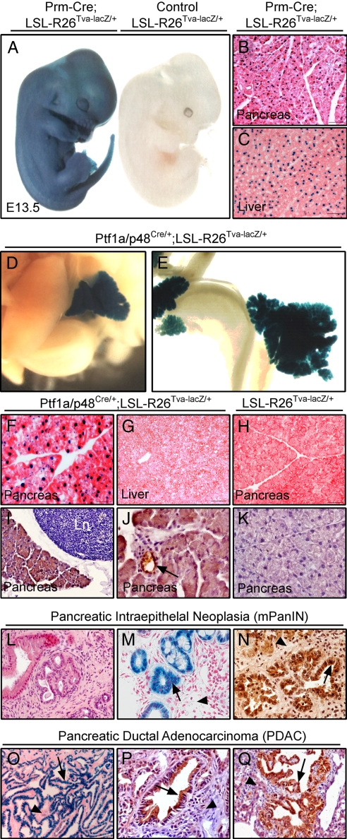
Fig. 1.
Characterization of LSL-R26Tva-lacZ knockin mice. (A) Whole-mount X-Gal staining of E13.5 LSL-R26Tva-lacZ/+;Prm-Cre (Left) and LSL-R26Tva-lacZ/+ (Right) embryos. (B and C) Nuclear lacZ activity in sections of the pancreas (B) and liver (C) isolated from adult LSL-R26Tva-lacZ/+;Prm-Cre mouse. (D–G) Visualization of lacZ activity in LSL-R26Tva-lacZ/+;Ptf1a/p48Cre/+ mice. Macroscopic images of X-Gal-stained liver and pancreas (D) and small bowel and pancreas (E) of E18 embryo. Microscopic images of nuclear lacZ activity in sections of the pancreas (F) and the liver (G) of adult mouse. (H) X-Gal staining reveals no lacZ activity in pancreatic sections of adult LSL-R26Tva-lacZ/+ mouse (control). (I–K) Immunostaining for TVA (brown color) in the pancreas of adult LSL-R26Tva-lacZ/+;Ptf1a/p48Cre/+ (I and J) and control LSL-R26Tva-lacZ/+ (K) mouse. TVA is expressed in islets (data not shown), ducts (black arrow in J) and acini (I and J), but not adjacent lymph node (Ln in I). (L–N) Expression of lacZnls and TVA in mPanIN lesions of Ptf1a/p48Cre/+;LSL-R26Tva-lacZ/+;LSL-KrasG12D/+ animals. Hematoxylin and eosin (H&E) (L), X-Gal (M) and immunohistochemical TVA (N) staining of pancreatic sections showing expression of nuclear lacZ (M) and TVA (N) in mPanIN lesions (black arrows) but not desmoplastic stroma (black arrowheads). (O–Q) Expression of TVA and lacZnls in murine PDAC of Ptf1a/p48Cre/+;LSL-R26Tva-lacZ/+;LSL-KrasG12D/+;LSL-TP53R172H/+ animals. X-Gal (O) and immunohistochemical TVA (P and Q) staining of sections from primary PDAC (O and P) and liver metastases (Q) showing expression of nuclear lacZ and TVA in PDAC (black arrow) but not desmoplastic stroma (O and P; black arrowheads) or adjacent normal liver (Q; black arrowhead).

