
Figure 4. MAGP-2 regulates Notch signaling in a cell type specific manner.
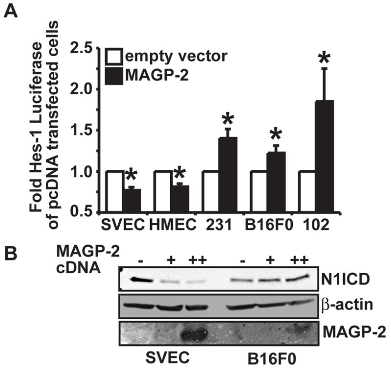
(A) Endothelial cells lines (SVEC and HMEC) and various tumor cell lines (MDA-MB-231 (231) breast carcinoma cells, B16F0 melanoma cells (B16), and MCA102 fibrosarcoma cells (102)) were transiently transfected with either empty vector (pcDNA3.1 myc-his) or MAGP-2 cDNA (pcDNA3.1-MAGP-2-myc-his) together with pHes-1 luciferase and pCMV-β-gal. Afterward, luciferase and β-gal activities were measured as above. Data are the mean (± SEM) of three independent experiments. (*, p < 0.05; Student’s Test). (B) SVEC ECs and B16F0 melanoma cells were transiently transfected with either empty vector (−) or with 1 (+) or 2.5 (++)μg of MAGP-2 cDNA and endogenous Notch1 NICD fragments were monitored by western blot analysis of whole cell lysates with anti-Val1744 antibodies. The western blot was subsequently stripped and reprobbed with anti-β-actin antibodies to monitor differences in protein loading. Conditioned media from the transfected cells was collected, precipitated with TCA, and analyzed by western blot with anti-MAGP-2 antibodies to monitor MAGP-2 expression. Shown is a representative experiment that was performed three times in its entirety with similar results.