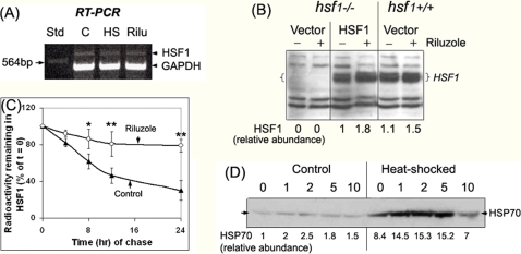
Figure 6. Effects of riluzole on the expression, turnover and activity of HSF1.
(A) Reverse-transcriptase PCR analysis of the effects of heat shock and riluzole treatment on the expression of mRNAhsf1. RNA was isolated from control, heat shocked (2 hr, 42°C), and riluzole-treated (2 μM, 8 hr) HeLa cells. RNA was reverse transcribed and PCR-amplified using HSF1-specific primers. Std: a 564 bp fragment from HindIII digest of λ DNA. The positions of the 642 bp HSF1 DNA fragment and the co-amplified 481 bp GAPDH internal control are as indicated. (B) Effects of riluzole on the endogenous versus CMV-promoter driven HSF1 expression. An episomal eukaryotic expression vector of the human HSF1, pCep4hHSF1, was used to drive the expression of HSF1 in hsf1−/− MEF. Cells were allowed to recover at 37°C for 6 hr after DNA transfection. Riluzole was then added to designated plates to a final concentration of 2 μM and incubated at 37°C for 16 hr. Cells were harvested and RIPA extracts prepared. Control and riluzole-treated hsf1−/− cells transfected with the pCep4 vector as well as hsf+/+ cells were included as controls in the experiment. Result on the relative abundance of HSF1 is shown at the bottom of the figure. (C) Pulse-chase analysis of the turnover of HSF1 in control versus riluzole-treated HeLa cells. Cells were labeled with [35S]methionine as described in the text. At the end of this labeling period, cells were rinsed extensively the chase initiated either without (Control, solid symbol) or with 2 μM riluzole at 37°C (open symbol). Samples were harvested at 0, 4, 8, 12 and 24 hr after initiation of the chase. Aliquots of the cell extracts were used for immunoprecipitation of HSF1 using protein A for pull-down by centrifugation. Result on the amount of radioactivity remaining with the HSF1 immunoprecipitate, relative to that of the t = 0 chase control, is plotted against the time of chase. Result is the average of 4 separate determinations±standard deviation. The single and double asterisk symbols, * and **, indicate two tailed t-test of the control vs. riluzole-treated samples with probability of difference of 0.01–0.05 (*, significant) and <0.01 (**, highly significant), respectively. (D) Dose-response effect of riluzole on HSP70 expression under control and heat-shocked conditions. HeLa cells were treated with concentrations of riluzole as indicated for a total of 18 hrs. For heat shock, cells, at 10 hrs after the addition of riluzole, were placed in a 42°C incubator for 2 hr followed by recovery at 37°C for 6 hr. Aliquots of the RIPA cell extract containing 10 μg protein were used for immuno-Western blot analyses of HSP70 [23]. The relative abundance of HSP70 is indicated at the bottom of the figure.

