
Figure 2.
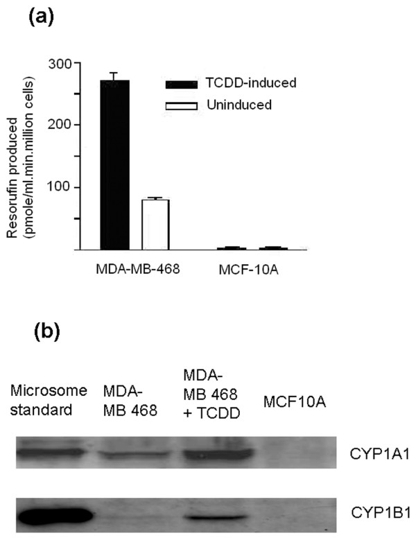
CYP1 enzyme expression in MDA-MB-468 and MCF-10A cells. (a) EROD activity of MDA-MB-468 and MCF-10A cells. Cells were seeded at a density of 5 × 10⁴ cells per millilitre in 24-well plates and left to grow for 48 hours. EROD activity was measured as described in Materials and methods. Error bars represent mean ± standard deviation for n = 4 determinations. (b) Selective and inducible CYP1A and CYP1B1 expression in MDA-MB-468 cells. Lysates were probed with anti-CYP1A and anti-CYP1B1 antibodies from Gentest Corporation (now part of BD Biosciences) and Auvation Limited. Lane 1: Recombinant CYP1A1 (top, 0.2 μg) or CYP1B1 (bottom, 0.4 μg) used as positive control. Lane 2: MDA-MB-468 cells. Lane 3: MDA-MB-468 cells treated with 10 nM TCDD for 24 hours. Lane 4: MCF-10A cells. Experiments were performed in duplicate. EROD, ethoxyresorufin-O-deethylase; TCDD, 2,3,7,8-tetrachlorodibenzo-p-dioxin.