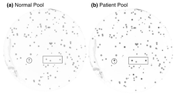
Figure 1.

Identification of disease-specific phage clones after the biopanning process. Two nitrocellulose membrane disks were placed on and then lifted from the same phage grown plate of biopan 4. (a) One membrane was probed with pooled normal sera and (b) the other was probed with pooled patient sera. After electrogenerated chemiluminescence detection, numerous immunoreactive clones showed more intensified spots on the membrane incubated with patient sera than on the membrane incubated with normal sera. The circle and square indicate the same area on the two membranes.
