
Figure 1.
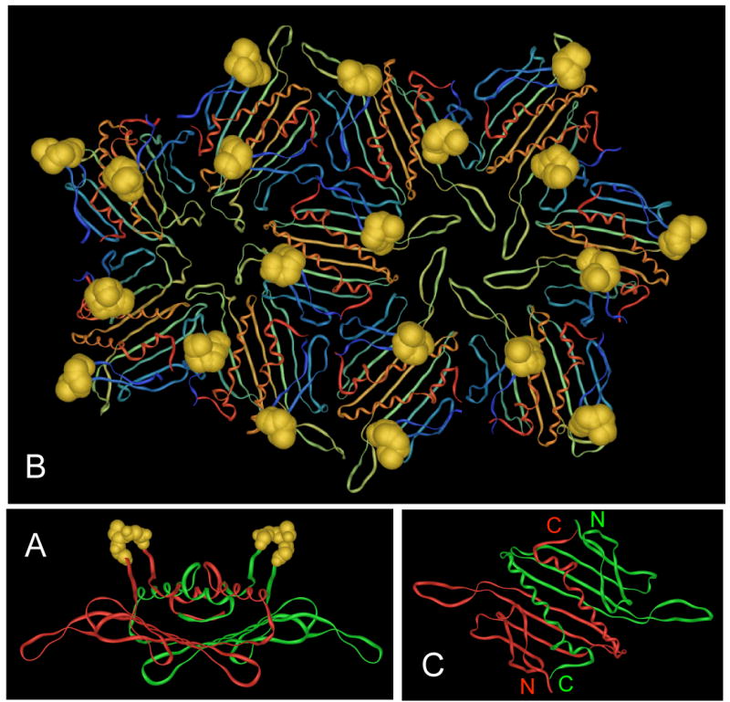
(A) Structure of the MS2 coat protein dimer seen edge-on, with its polypeptide chains in red and green and the AB-loops in gold. (B) A portion of the MS2 VLP showing the arrangment of subunits around the 5-fold and quasi-6-fold symmetry axes and the arrangement of the AB-loops (in gold). (C) Structure of the MS2 coat protein dimer as viewed from outside the VLP. Note the proximity of the N- and C-termini of the two polypeptides. Images were produced using imol software, available at http://www.pirx.com/iMol/.