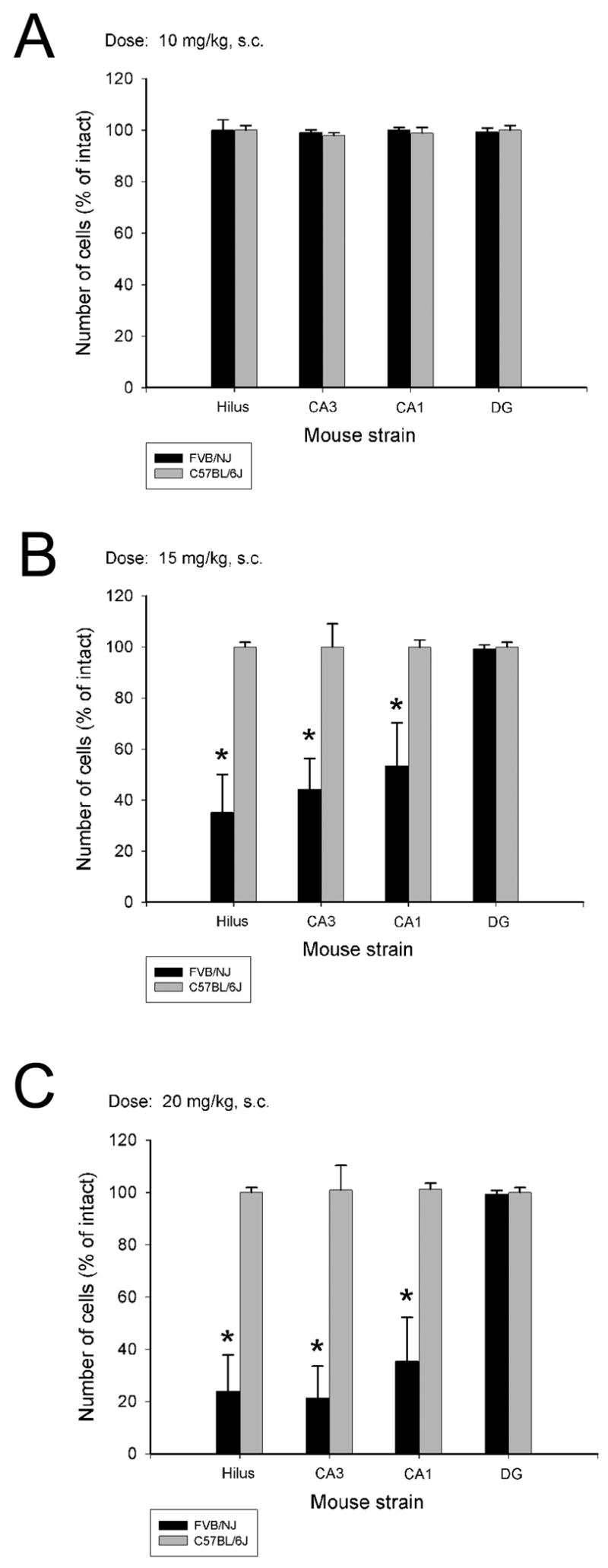
Fig. 3.

Quantitative analysis of neuronal density in hippocampal subfields following administration of 3 different doses of KA to middle-aged C57BL/6 and FVB/NJ mice. While no cell loss as observed at the lowest dose of KA (A), a significant increase in cell loss was observed in the dentate hilus, and areas CA3 and CA1 of FVB/NJ mice seven days following KA administration. Data represent the mean ± SEM of at least 8 mice/strain. *P<0.05.
