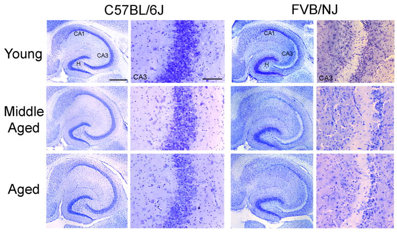
Fig. 5.
Neuronal cell loss following kainate administration in C57BL/6J and FVB/NJ mice is strain-dependent in three different age groups of mice. Low- and high-power photomicrographs of cresyl violet-stained horizontal sections of the hippocampus showing the destruction of neurons in the CA3 and CA1 subfields and within the dentate hilus 7 days after kainate administration in all three age groups of FVB/NJ mice. In contrast, no cell loss was evident in C57BL/6J mice of any age group following kainate administration. CA3, CA3 pyramidal cell layer; CA1, CA1 pyramidal cell layer; H, hilus. Scale bars, 750 μm (low-power photomicrographs); 350 μm (high-power photomicrographs).

