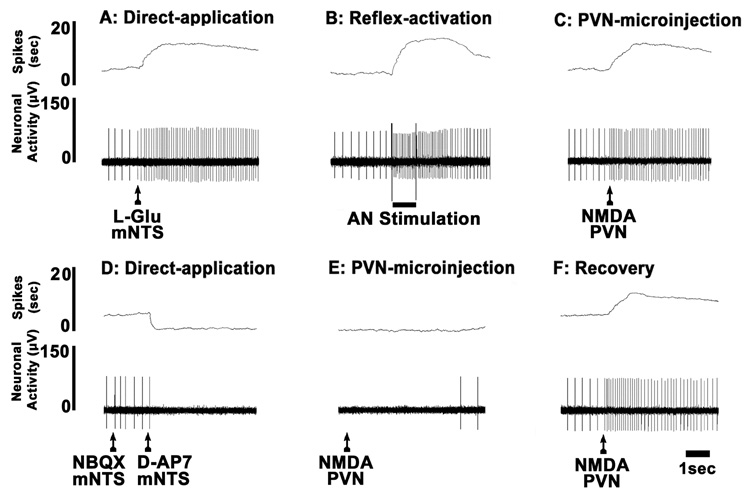
Fig. 4.
A typical tracing of extracellular neuronal recording in a barodenervated rat. The firing of the mNTS neuron was increased by direct application of L-Glu (5 mM, 4 nl) (A), electrical stimulation of the ipsilateral aortic nerve (B), and microinjection of NMDA (10 mM, 20 nl) into the ipsilateral PVN (C). Direct application of NBQX (2 mM, 4 nl) and D-AP7 (5 mM, 4 nl) abolished the neuronal firing (D). Within 10 sec, NMDA (10 mM, 20 nl) was again microinjected into the PVN; no excitation was observed due to ionotropic glutamate receptor blockade of the mNTS neuron (E; compare with C). After 5 min, NMDA (10 mM, 20 nl) was again microinjected into the PVN; the excitation of the neuron resumed (F, compare with C).

