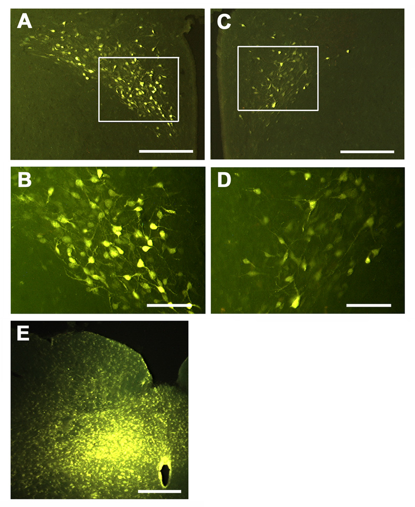
Fig. 6.

Retrograde labeling of PVN neurons. A, C and E = low magnification. B and D high magnification of boxed areas in A and C, respectively. Labeling of ipsilateral (A) and contralateral (C) PVN following unilateral microinjection (15 nl) of fluorogold (4–6%) into the mNTS (E). Examination of the section shows preponderance of the labeling in the ipsilateral PVN (A and B). Scale bars: 300 µm in A and C; 100 µm in B and D; 400 µm in E.
