
Fig. 7.
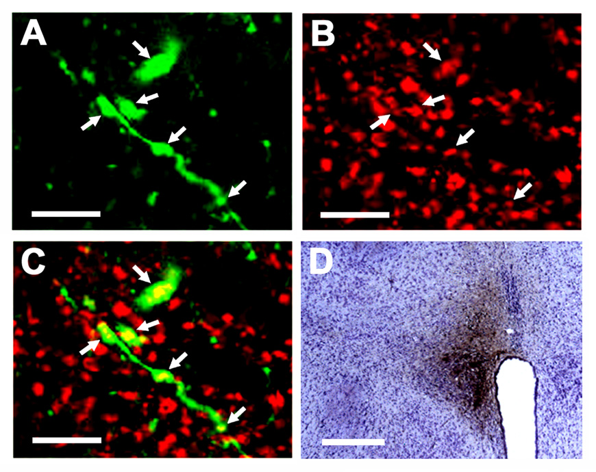
Immunoreactivity of vesicular glutamate transporter-2 (VGLUT2) in the terminals of PVN neurons innervating the mNTS. A cluster of BDA-labeled axons and terminals (green) is shown in panel A (confocal microscopy; Alexa Fluor 488 fluorescence). VGLUT2 immunoreactivity (red) in the same field is shown in panel B (Cy3 fluorescence). A merged image shown in panel C indicates that the BDA-labeled terminals are VGLUT2-immunoreactive (colocalization is shown by yellow color). The site of microinjection of BDA (10%, 100 nl) in the PVN is shown in panel D. Scale bars; 20 µm (panels A, B, C) and 300 µm (panel D).