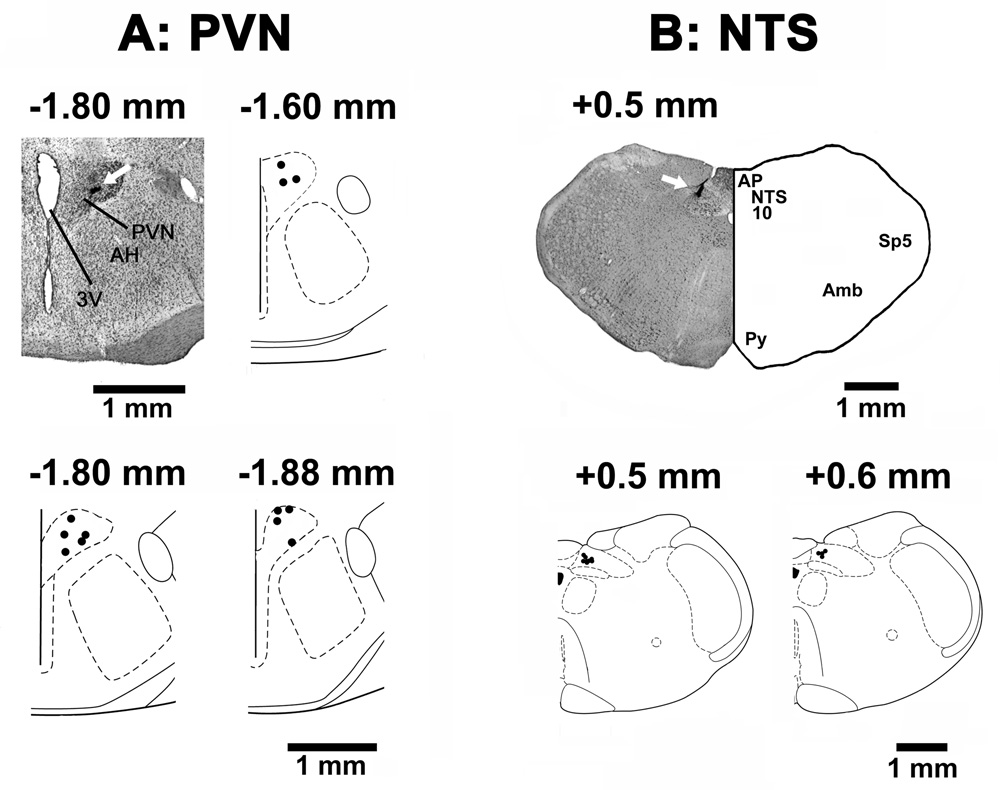
Fig. 8.
Histological identification of microinjection sites. A: A coronal section at a level 1.8 mm caudal to the bregma showing a typical PVN microinjection site marked (arrow) with India ink; the center of the spot was 0.5 mm lateral to the midline and 7.8 mm deep from the dura. Composite diagrams in this panel represent the PVN sites (n = 12), where NMDA was microinjected, at levels 1.60, 1.8 and 1.88 mm caudal to the bregma. B: A coronal section at a level 0.5 mm rostral to the calamus scriptorius showing a typical mNTS microinjection site marked (arrow) with India ink; the center of the spot was 0.5 mm lateral to the midline and 0.5 mm deep from the dorsal medullary surface. Composite diagrams in this panel represent the mNTS sites (n = 12) at levels 0.5 and 0.6 mm rostral to the calamus scriptorius. In the composite diagrams, each spot represents the microinjection site in one rat; all spots are not visible due to some overlap. Abbreviations: AH: anterior hypothalamic nucleus; Amb: nucleus ambiguus, AP: area postrema, NTS: nucleus tractus solitarius; PVN: hypothalamic paraventricular nucleus; Py: pyramids; Sp5: spinal trigeminal tract; 3V: 3rd ventricle; 10: dorsal motor nucleus of vagus.

