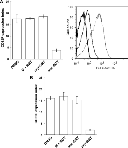
Figure 5.
Effect of myr-RGT on platelet CD62P expression in the presence of agonists. The expression of CD62P on nontreated or peptide-treated platelets stimulated with agonists was analyzed by flow cytometry using an FITC-labeled monoclonal anti-CD62P antibody. (A) Data shown in the left pattern (mean ± SD) were derived from the ratio of the geometric mean fluorescence intensity measured for anti-CD62P antibody binding to thrombin-treated platelets, preincubated for 30 minutes with DMSO vehicle, nonmyristoylated RGT peptide (250 μM) plus myristic acid (M + RGT), scrambled myr-GRT (250 μM), or myr-RGT (250 μM), versus resting platelets (without thrombin treatment) and obtained from 3 separate experiments. Data shown in the right pattern are a representative figure of CD62P expression in the presence of thrombin on platelets preincubated with DMSO (fine line), myr-RGT (normal line), or on resting platelets (in the absence of thrombin; thick line). (B) Expression of CD62P on peptide-treated platelets stimulated with ADP under stirring conditions.

