Figure 1.
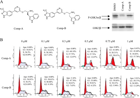
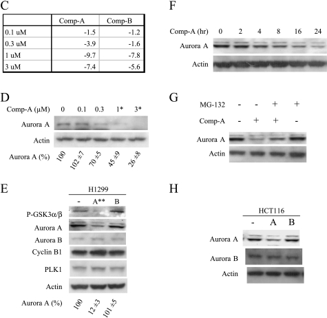
Akt inhibition down-regulates Aurora A and induces mitotic arrest. (A) Western analysis of P-GSK3α/β and total GSK3α/β in H1299 cells treated with Compound A or B at 0.6 µM concentration for 2 hours. (B) FACS analysis of H1299 cells treated with Compound A or B for 24 hours. (C) MiaPaca-2 cells were treated with Compound A or B at the indicated concentrations for 5 hours. Total RNA was isolated and subjected to microarray analysis as described. The fold changes compared to the DMSO control in Aurora A mRNA levels were listed. (D) MiaPaca-2 cells were treated with Compound A at the indicated concentrations for 24 hours. The cells were harvested, and Western blot analysis was carried out. Aurora A levels were quantified from three experiments using GS-800 calibrated densitometer (Bio-Rad, Hercules, CA) and listed beneath each western gels. Student's t test were performed and *P < .05 or **P < .01 is obtained between the control and the marked lane. (E) H1299 cells were treated with Compound A or B at 0.6 µM for 24 hours. The cells were harvested, and Western analysis was carried out. Statistical analysis was done as in (D). (F) H1299 cells were treated with Compound A at 0.6 µM for the indicated times. The cells were harvested, and Western analysis was carried out. (G) H1299 cells were treated with Compound A at 0.6 µM in the presence or absence of 20 µM MG132 for 24 hours. The cells were harvested, and Western analysis was carried out. (H) HCT116 cells were treated with Compound A or B at 0.5 µM for 24 hours. The cells were harvested, and Western analysis was carried out.


