Figure 6.
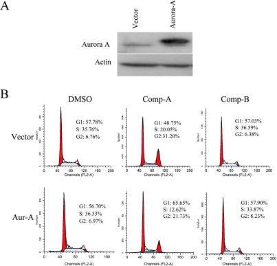
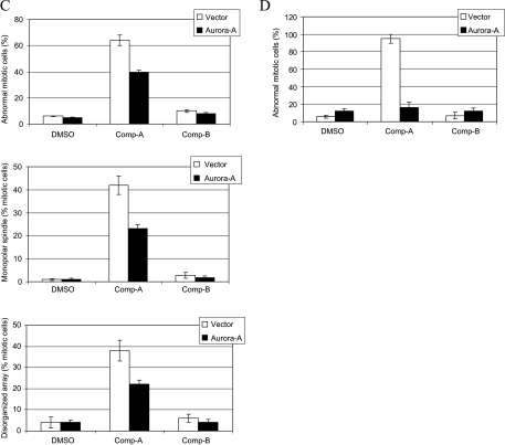
Overexpression of Aurora A rescues the mitotic defects induced by Akt inhibition. (A) H1299 cells were transfected with pcDNA3.1 or pcDNA3-Aurora A. Cell extracts were prepared 24 hours after transfection and were subjected to Western blot analysis. (B and C) H1299 cells were transfected as described in (A). The cells were then treated with DMSO, 0.6 µM Compound A or B for 24 hours. Cells were stained with propidium iodide for FACS analysis or were immunostained with DAPI and antibodies against α-and γ-tubulins. FACS analysis (B) and abnormal mitosis (C) were scored as described. For each condition, more than 150 mitotic cells were scored. Data are average of three independent experiments. (D) Experiment was done as in (C) except 0.8 µg of padtrack-GFP was cotransfected with pcDNA3.1-Aurora A. GFP were immunostained with fluorescein isothiocyanate-conjugated GFP antibody (Abcam, Cambridge, MA). Only GFP-positive cells (60–100 cells) were scored.


