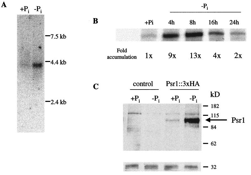
Figure 3.
Northern blot hybridization, RNase protection assays, and Western blot analysis of the Psr1 protein. (A) Northern blot hybridization of a Psr1 gene-specific probe to RNA from the wild-type strain CC125. Polyadenylated RNA from cells grown in nutrient-replete medium (+Pi) and from cells starved for 8–48 h and pooled (−Pi) was analyzed. (B) RNase protection of a 395-base riboprobe by 30 μg of total RNA. The level of accumulation of the transcript (fold change relative to +Pi conditions) is indicated beneath each lane. This experiment was repeated on separately isolated RNA samples, and the trend was identical. (C) Western blot analysis of the Psr1 protein containing the 3×HA tag. The first two lanes contain protein that was extracted from cells in which Psr1 did not contain the 3×HA tag. The second two lanes contain protein that was extracted from the psr1–1 mutant containing the PSR1∷3×HA construct. Protein extracts were from either cells grown in complete medium (+Pi) or cells starved for P for 24 h (−Pi). Three independent transformants were examined, and all three gave essentially identical results. To control for loading, the blot was stripped and reprobed for D1 protein (32-kDa polypeptide), which was shown previously not to change in abundance after 24 h of phosphorus starvation (6).

