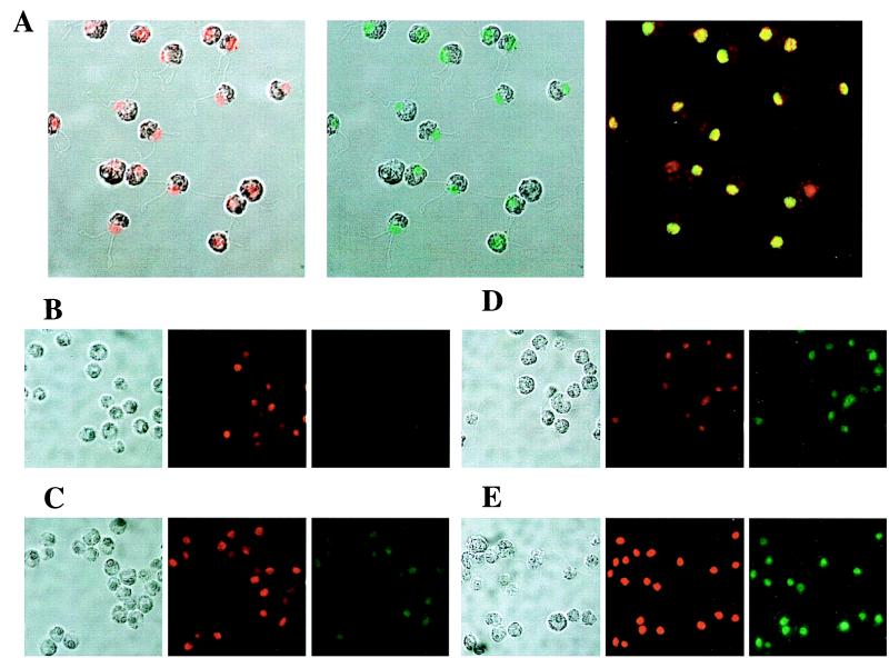
Figure 4.
Immunolocalization of Psr1∷3xHA. (A) Localization of DNA (red with transmission overlay, Left), Psr1∷3×HA (green with transmission overlay, Center), and both DNA and Psr1∷3×HA (yellow is coincident localization, Right). (B– E) Transmission microscopy (Left) after staining with propidium iodide for DNA (red, Center), or after incubation with antibodies to the HA tag and the fluorescent Alexa 488-anti rat conjugate (green, Right). B is a psr1 mutant complemented with the wild-type copy of Psr1 and starved for P for 24 h. C is the Psr1∷3×HA strain grown under nutrient replete conditions. D is the Psr1∷3×HA strain starved for P for 8 h. E is the Psr1∷3×HA strain starved for P for 24 h.

