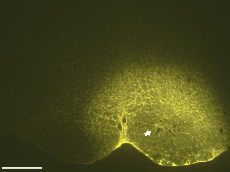
Fig. 1.

A photomicrograph (4X) of the FluoroGold injection site that resulted in retrograde labeling seen in Fig. 2 and Fig. 3. The center of the injection, characterized by a compacted core, lies dorsal to the pyramid and includes both the nucleus raphe magnus and the nucleus reticularis gigantocellularis pars alpha. A halo of neurons surrounds the core and represents local neurons that have been labeled by the tracer. Scale bar = 500 µm; distance from bregma approximately −11.60 mm.
