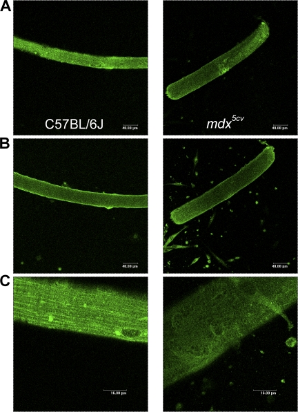
Figure 6.
Distribution pattern of Nav1.4 is altered in mdx fibers. Nav1.4 channel distribution in isolated FDB fibers; comparison between C57BL/6J and mdx5cv (four mice each condition). Nav1.4 immunofluorescence (green) in single C57BL/6J fibers (left) and mdx5cv fibers (right) captured at the surface (A) or in deeper part of the cells (B). (C) 4× zoom of A.

