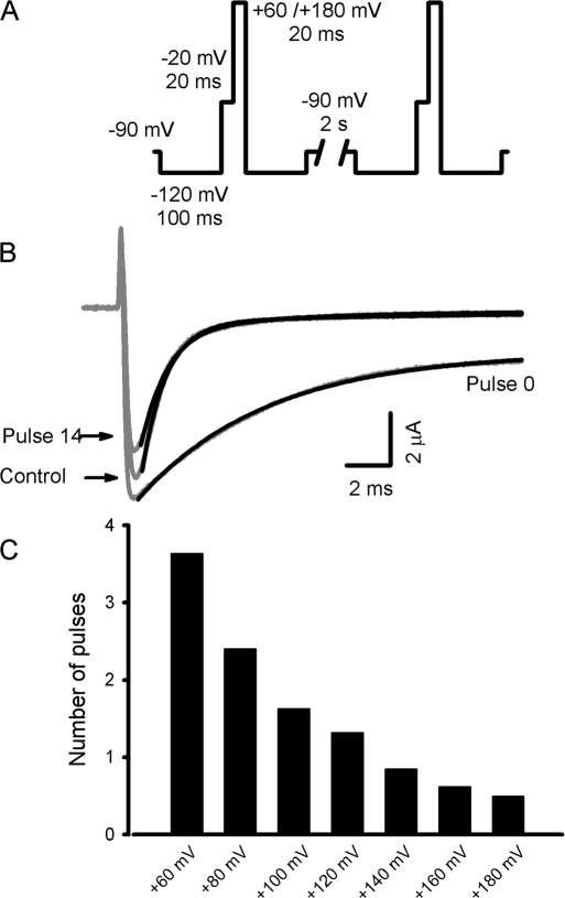
Figure 2.
Ts3 voltage-dependent displacement. (A) Pulse protocol used to remove the bound Ts3. A strong depolarizing pulse, varying from +60 to +180 mV during 20 ms, was applied just after a −20 mV test pulse. Between pulses the oocytes were held at −90 mV, and the protocol was repeated at least 15 times. (B) Traces (gray lines) obtained in control conditions, in the presence of Ts3 (Pulse #0) and after 14 successive depolarization to +120 mV, by applying the protocol described above (Pulse #14) in the absence of Ts3. Black lines shows the curves obtained by fitting the data with function 1 (see Materials and methods). (C) Voltage dependence of toxin removal, obtained by applying the pulse protocol described in A. The graph shows the data obtained from a representative experiment. The number of pulses needed to an e-fold displacement was calculated by fitting the slow component contribution decay with a single exponential function.

