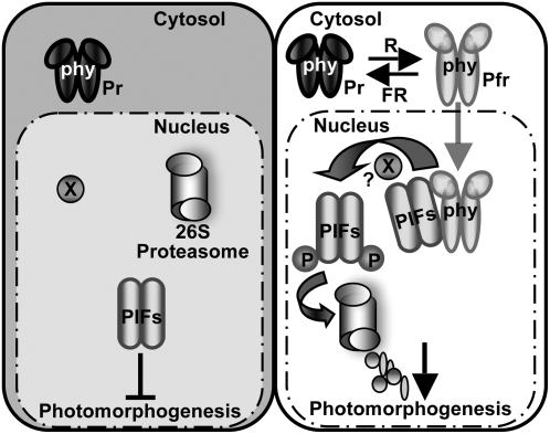
Figure 12.
Simplified Model of PIF Function in phy Signaling Pathways.
Left, in the dark, phys are localized to the cytosol, while PIFs are constitutively localized to the nucleus and negatively regulate photomorphogenesis. Right, light signals promote nuclear migration of phys by inducing the photoconversion of the Pr form to the active Pfr form. In the nucleus, the photoactivated phys interact with PIFs, resulting in the phosphorylation of PIF1 and other PIFs either directly or indirectly. The phosphorylated forms of PIFs are then polyubiquitinated by a ubi ligase and subsequently degraded by the 26S proteasome. The light-induced proteolytic removal of PIFs relieves the negative regulation, thus promoting photomorphogenesis. X indicates an unknown factor that might be involved in the light-induced phosphorylation of PIFs. P, phosphorylated form. This figure is adapted and modified from Castillon et al. (2007).

