Figure 3.
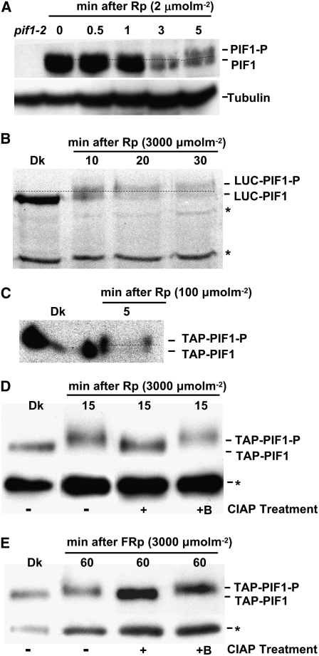
Light Induces Rapid Phosphorylation prior to Degradation of PIF1.
(A) Native PIF1 migrates as two bands (PIF1 and PIF1-P) following Rp (2 μmol·m−2). A blot probed with anti-PIF1 antibody is shown.
(B) LUC-PIF1 also exhibits a slower migrating band (LUC-PIF1-P) after Rp (3000 μmol·m−2). Proteins from plants expressing LUC-PIF1 were probed with anti-LUC antibody.
(C) TAP-PIF1 shows a slower migrating band (TAP-PIF1-P) and is also degraded after Rp (100 μmol·m−2). Proteins from plants expressing TAP-PIF1 were probed with anti-MYC antibody that recognizes the TAP tag.
Dotted lines separate the two forms of PIF1 in (A) to (C).
(D) and (E) The Rp- and FRp-induced slow-migrating band is a phosphorylated form of PIF1. TAP-PIF1 was immunoprecipitated from protein extracts prepared using 4-d-old dark-grown 35S:TAP-PIF1 seedlings kept in the dark or exposed to either Rp (3000 μmol·m−2; [D]) or FRp (3000 μmol·m−2; [E]) followed by dark incubation. The immunoprecipitated pellets from the Rp- or FRp-exposed samples were dissolved in buffer and incubated without (−) or with (+) native CIAP or with boiled CIAP (+B). Samples were then separated on 6.5% SDS-PAGE gels and probed with anti-MYC antibody. Asterisks denote cross-reacting bands.

