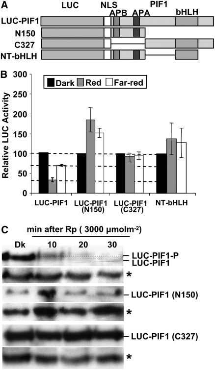
Figure 8.
Both the N and C Termini of PIF1 Are Necessary for the Light-Induced Degradation of PIF1.
(A) Design of the PIF1 deletion constructs fused to LUC. The white boxes represents a nuclear localization signal (NLS).
(B) LUC activity was measured from 4-d-old dark-grown seedlings transferred to R (10 μmol·m−2·s−1) or FR (10 μmol·m−2·s−1) light for 1 h as described (Shen et al., 2005). Means ± se of five biological replicates are shown. Some constructs showed greater stability of the fusion protein in light relative to darkness for unknown reasons.
(C) Protein gel blots showing truncated PIF1 fusion proteins are neither phosphorylated nor degraded under light, but the wild-type LUC-PIF1 is both phosphorylated and degraded under light. The dotted line separates the two forms of PIF1. Asterisks denote a cross-reacting band.

