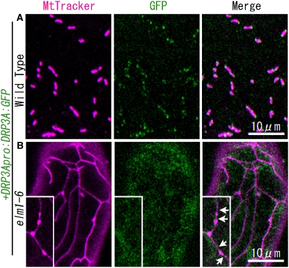
Figure 5.
DRP3A:GFP Does Not Localize to Mitochondrial Ends and Constricted Sites in the elm1-6 Mutant.
Confocal laser scanning microscopy images of the cotyledon leaf epidermal cells of wild-type plants (A) and elm1-6 T-DNA insertion mutants (B) transformed with the construct DRP3Apro:DRP3A:GFP. Mitochondria were visualized by MitoTracker Orange staining. Insets in (B) show beads-on-a-string-shaped mitochondria that transiently appeared in elm1-6 from a separate microscope field. Arrows show the constricted sites on mitochondria in elm1-6. Bar in the bottom panel indicates scale of insets.

