
Fig. 1.
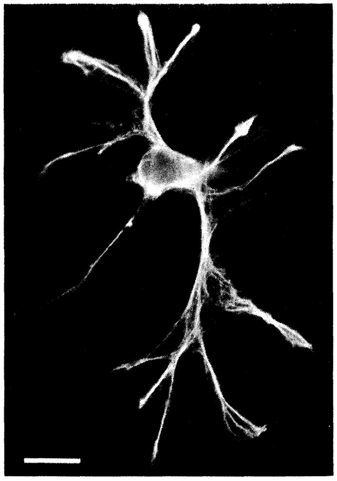
GFAP immunoreactivity of a freshly dissociated cell from the optic nerve of the salamander (19). This fluorescence micrograph shows that the cell was heavily labeled with antibodies to GFAP, identifying it unequivocally as an astrocyte. Scale bar, 20 μm.