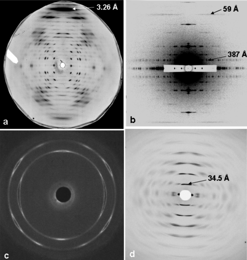
Figure 1.
Sample diffraction patterns from non-crystalline materials. (a) A well aligned polycrystalline specimen of E-DNA [poly(I-I-T).poly(A-C-C) (from Leslie et al., 1980 ▶)]. (b) A low-angle X-ray diffraction pattern from insect flight muscle, showing excellent sampling and d spacings in the range 48–387 Å with a c-axis repeat of 2320 Å (from AL-Khayat et al., 2004 ▶). (c) Wide-angle X-ray diffraction pattern from a sample of a diblock copolymer of oxyethylene (E) and oxybutylene (B) as E76B38 (where the subscripts denote the average numbers of repeat units) that had been crystallized after shearing in the melt (Fairclough et al., 2000 ▶). (d) High-angle X-ray diffraction pattern from magnetically oriented sols of PotatoVirus X, showing a helical repeat of about 34.5 Å (Stubbs et al., 2005 ▶).

