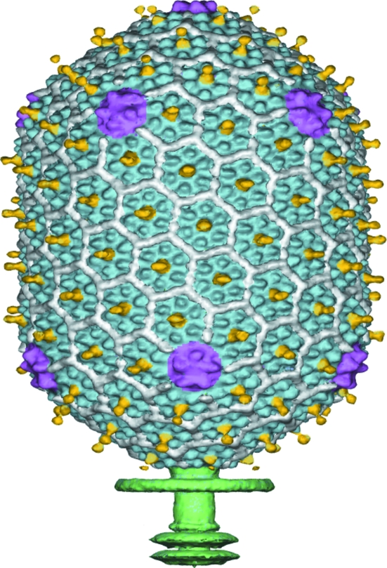
Figure 5.
Cryo-EM reconstruction of the head capsid of bacteriophage T4, based on fivefold symmetry averaging. The major capsid protein (gp23, in blue) forms hexamers. The small outer capsid protein (soc, in white) binds between gp23 hexamers. The highly antigenic outer capsid protein (hoc, in yellow) binds at the center of gp23 hexamers. Pentamers of the special vertex protein gp24 (purple) are at the icosahedral vertices. The tail (green) is smeared as it has sixfold symmetry, not the fivefold symmetry used for averaging.

