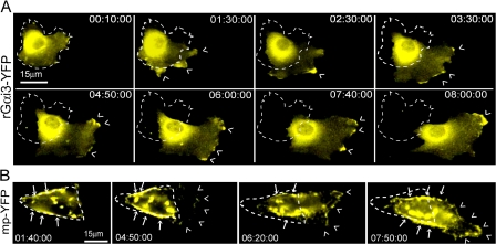
Figure 2.
Gαi3 localizes preferentially in pseudopods at the leading edge. (A) Time-lapse images of a HeLa cell expressing Gαi3-YFP migrating into a freshly made wound visualized by live cell imaging. Gαi3-YFP is preferentially localized within pseudopods at the leading edge (carats) during polarized migration. Time after wounding is expressed as hours:minutes:seconds. Outline of the cell at the beginning of Video 2 (available at http://www.jcb.org/cgi/content/full/jcb.200712066/DC1) is indicated with a dashed line. (B) Similar images of a HeLa cell expressing mp-YFP. Mp-YFP shows a patchy distribution over most of the PM (arrows) and other membranous compartments with no preferential localization to the leading edge (carats). The dashed line indicates the position of the cell at the beginning of Video 3 (available at http://www.jcb.org/cgi/content/full/jcb.200712066/DC1).

