Figure 4.
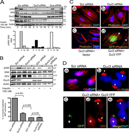
Gαi3 cooperates with GIV in Akt activation and actin remodeling and regulates the distribution of GIV. (A) Depletion of Gαi3 but not Gαs inhibits insulin-stimulated Akt activation. HeLa cells treated as indicated were serum starved for 6 h and stimulated with 100 nM insulin. (Top) Lysates were separated by SDS-PAGE and immunoblotted for total (tAkt), phospho-Akt (pAkt), Gαi3, Gαs, and actin. (Bottom) Bar graph showing ratio of pAkt to tAkt in scr RNA controls versus cells depleted of Gαi3 or Gαs. (B) Simultaneous depletion of Gαi3 and GIV does not have an additive effect on Akt signaling. (Top) HeLa cells treated as in A were immunoblotted for tAkt and pAkt 5 min after insulin stimulation. (Bottom) Bar graph showing Akt activity in cells treated as in top expressed as percentage of the response in controls. Results are shown as mean ± SEM (n = 3). P values compared with scr-siRNA are indicated. (C) Depletion of Gαi3 alters actin organization. Cells treated with scr siRNA (a) show stress fibers whereas Gαi3-depleted cells (b) are unable to form stress fibers and show a prominent bed of cortical actin. The effects on actin in Gαi3-depleted cells are reversed by transfecting rGαi3wt-YFP (d) but not vector alone (c). HeLa cells treated as indicated were stained with Phalloidin (red) and DAPI (blue). *, cells expressing rGαi3-YFP visualized with anti-GFP. Bar, 10 μm. (D) Distribution of GIV is less scattered and more concentrated in the Golgi (arrowheads) upon silencing Gαi3 (compare a and b). The scattered distribution is restored upon transfecting rGαi3-YFP (c–e). HeLa cells were treated as indicated and stained for GIV (red), GFP (green), and DAPI (blue). *, cell expressing rGαi3-YFP visualized with anti-GFP mAb. Bar, 10 μm.

