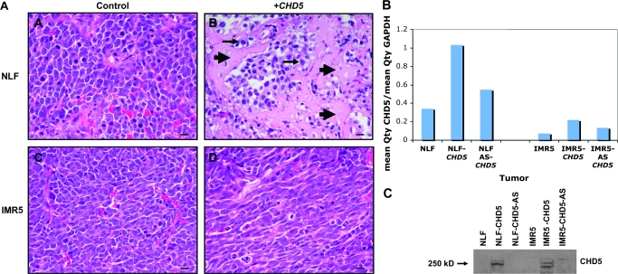
Figure 4.
Histology and CHD5 expression in xenografts derived from NLF and IMR5 cells transfected with either CHD5 sense or CHD5 antisense constructs. A) Histology of NLF and IMR5 xenograft tumors (see Figure 3) after 5 weeks of growth. NLF-CHD5-AS tumors were composed of undifferentiated cells with scant cytoplasm (top left), whereas NLF-CHD5 tumors showed areas of necrosis (arrowheads) and differentiation (arrows; top right). Cells in the IMR5-CHD5-AS tumors were undifferentiated (bottom left), whereas cells in the IMR5-CHD5 tumors had a more elongated appearance (bottom right). Bar = 20 μm. B) Relative expression of CHD5, normalized to glyceraldehyde-3-phosphate dehydrogenase (GAPDH), was determined by real-time reverse transcription–polymerase chain reaction in parental NLF and IMR5 cell lines, as well as the CHD5 sense and CHD5 antisense transfected lines used for the xenograft experiments. The normalized values indicated by each bar graph represent the mean of three measurements. Replicate measurements were within 10% of the mean for each bar shown. C) Expression of CHD5 protein, as detected by immunoblotting, is shown for the NLF and IMR5 parental lines and the corresponding sense- and antisense-transfected cells used in these experiments.

