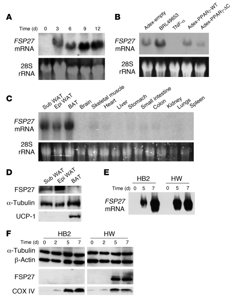
Figure 1. Adipocyte-specific expression of Fsp27.
(A) Northern blot analysis of FSP27 mRNA in 3T3-L1 cells at the indicated times after the onset of induction of adipocyte differentiation. The region of the ethidium bromide–stained gel containing 28S rRNA is also shown. (B) Northern blot analysis of FSP27 mRNA in 3T3-L1 adipocytes, either after incubation for 48 hours with 5 μM BRL49653 or TNF-α (10 ng/ml) or 48 hours after infection with adenoviral vectors encoding wild-type mouse PPARγ (adex-PPARγ-WT) or PPARγ-ΔC (adex-PPARγ-ΔC) or with the corresponding empty vector (adex-empty), at an MOI of 60 PFU per cell. (C) Northern blot analysis of FSP27 mRNA in various organs and tissues of C57BL/6J mice at 4 weeks of age. Sub, subcutaneous; Epi, epididymal. (D) Immunoblot analysis of FSP27 in total lysates of WAT (subcutaneous or epididymal) and BAT isolated from C57BL/6J mice at 4 weeks of age. Both α-tubulin (loading control) and UCP-1 (BAT marker) were also examined. (E) Northern blot analysis of FSP27 mRNA in HB2 and HW cells at the indicated times after the onset of induction of adipocyte differentiation. (F) Immunoblot analysis of FSP27 in HB2 and HW cells at the indicated times after the onset of induction of adipocyte differentiation using the antibodies to a COOH-terminal peptide of FSP27. Both α-tubulin and β-actin (loading controls) as well as COXIV (mitochondrial marker) were also examined.

