
Figure 9.
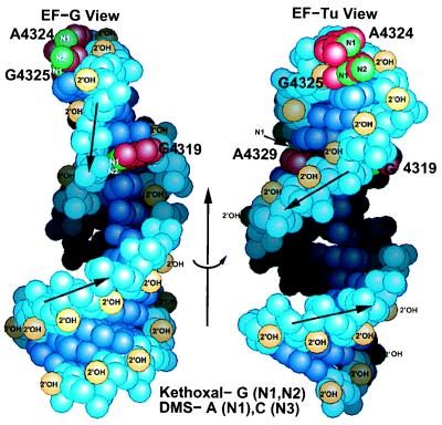
Mapping the chemical footprinting on the SRL structure. The left EF-G view is rotated 65° about the indicated arrow to generate the right EF-Tu view. The left drawing shows the unusual riboses of A4318 and G4319, which present their 2′-hydroxyls to the major instead of the minor groove. The structure is consistent with the chemical footprinting, since Watson–Crick faces of A4324, G4325, and A4329 are accessible. In the case of the bulged G4319, kethoxal is likely to approach perpendicular to the ring, since the 5′-side of G4319 is accessible and its Watson–Crick face is not.