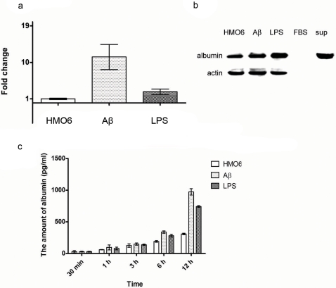
Figure 3. Increase in albumin synthesis and secretion after microglial activation by Aβ1–42- or LPS-treatments.
qRT-PCR data (a) show that the transcription of albumin gene is significantly enhanced after microglial activation by Aβ1–42- or LPS-treatment. Immunoblot data also show that albumin synthesis increases at the protein level after microglial activation (b). In addition, immunoblot data show that anti-human-albumin antibody used does not have any cross-reactivity with bovine albumin, and that albumin is present in the incubation medium (i.e. thus secreted from cells). ELISA data show that albumin secretion from HMO6 cells increased significantly after microglial activation (c). Moreover, the level of albumin in the incubation medium of Aβ1–42-treated cells was significantly higher than that of LPS-treated cells. This observation partly explains why the increase of albumin expression at the mRNA level is not reflected well at the protein level. Since albumin is directly secreted from cells after synthesis, the increase of albumin expression seems to be reflected better in the incubation media than in cell lysates.

