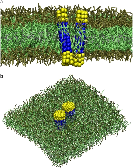
FIGURE 1.
Snapshot of the lipid bilayer with two embedded proteins of size Np = 7 (a) and Np = 43 (b). The hydrophilic and the hydrophobic beads of the proteins are depicted in yellow and in blue, respectively. The lipid headgroups are depicted in brown, the lipid tails in green and the terminal beads of the lipid tails in gray.

