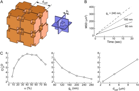
FIGURE 3.
Three-dimensional model of diffusion in brain ECS. (A) Schematic of three-dimensional model of diffusion in brain ECS showing a cubic lattice arrangement of cells containing lakes at multicell contact points (left). ECS geometry with narrow gaps and lake regions (right). (B) Representative MSD plots. Parameters α ∼ 21.5%, dcell = 10 μm, and indicated gc. (C) Do/D as a function of (left) α, (middle) gc, and (right) dcell. Parameters: (left) dcell = 10 μm, gc = 80 nm; (middle) α ∼ 21.5%, dcell = 10 μm; (right) α ∼ 21.5%, gc = 80 nm.

