
Figure 2.
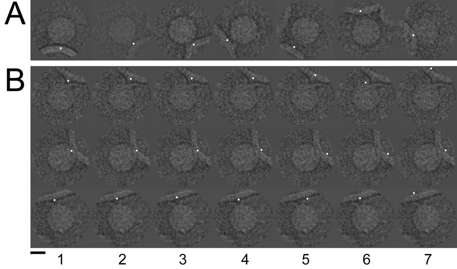
Cryo-electron microscope images of poliovirus-receptor-liposome complexes were artificially dotted to indicate the membrane contact point. The coordinate of this contact could be identified visually (A) as well as be defined by the final Eulerian angles reported for each image by Bubeck et al. 2005 (B, column 1). Column 2 illustrates the addition of random Gaussian error with an RMSD of 5 pixels. Columns 3–7 show error levels of 10, 20, 30, 40, and 50 RMSD. Scale bar, 150 Å.